Serine/threonine kinase which acts as an essential component of the MAP kinase signal transduction pathway. Plays an important role in the cascades of cellular responses evoked by changes in the environment. Mediates signaling for determination of cell fate such as differentiation and survival. Plays a crucial role in the apoptosis signal transduction pathway through mitochondria-dependent caspase activation. MAP3K5/ASK1 is required for the innate immune response, which is essential for host defense against a wide range of pathogens. Mediates signal transduction of various stressors like oxidative stress as well as by receptor-mediated inflammatory signals, such as the tumor necrosis factor (TNF) or lipopolysaccharide (LPS). Once activated, acts as an upstream activator of the MKK/JNK signal transduction cascade and the p38 MAPK signal transduction cascade through the phosphorylation and activation of several MAP kinase kinases like MAP2K4/SEK1, MAP2K3/MKK3, MAP2K6/MKK6 and MAP2K7/MKK7. These MAP2Ks in turn activate p38 MAPKs and c-jun N-terminal kinases (JNKs). Both p38 MAPK and JNKs control the transcription factors activator protein-1 (AP-1).
Description
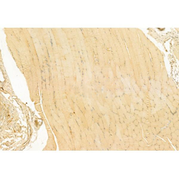
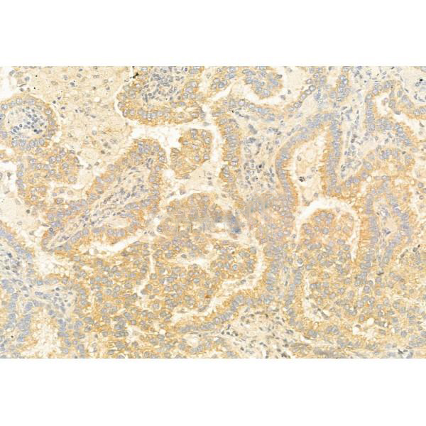
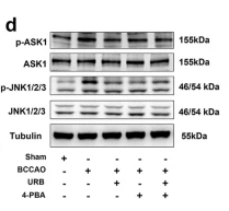
Rabbit polyclonal antibody to ASK1
Applications
WB, IF, ICC, IHC.
Immunogen
ASK1 Antibody detects endogenous levels of total ASK1.
Reactivity
Human, Mouse, Rat.
可预测:Pig(100%), Zebrafish(%), Bovine(%), Horse(%), Sheep(%), Rabbit(%), Dog(%), Chicken(%)
Molecular weight
155kDa; 155kD(Calculated).
Host species
Rabbit
Ig class
Immunogen-specific rabbit IgG
Purification
Antigen affinity purification
Full name
ASK1
Synonyms
Apoptosis signal regulating kinase 1; Apoptosis signal-regulating kinase 1; ASK 1; ASK-1; ASK1; M3K5; M3K5_HUMAN; MAP/ERK kinase kinase 5; MAP3K5; MAPK/ERK kinase kinase 5; MAPKKK5; MEK kinase 5; MEKK 5; MEKK5; Mitogen activated protein kinase kinase kinase 5; Mitogen-activated protein kinase kinase kinase 5;
Storage
Rabbit IgG in phosphate buffered saline , pH 7.4, 150mM NaCl, 0.02% sodium azide and 50% glycerol. Store at -20 °C. Stable for 12 months from date of receipt.
Swissprot
Q99683


 产品订购:
产品订购:
 渠道电话:
渠道电话: