Forms a water-specific channel that provides the plasma membranes of red cells and kidney proximal tubules with high permeability to water, thereby permitting water to move in the direction of an osmotic gradient.
Description
Rabbit polyclonal antibody to AQP1
Applications
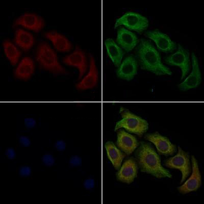
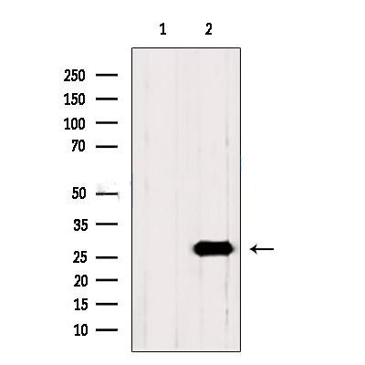
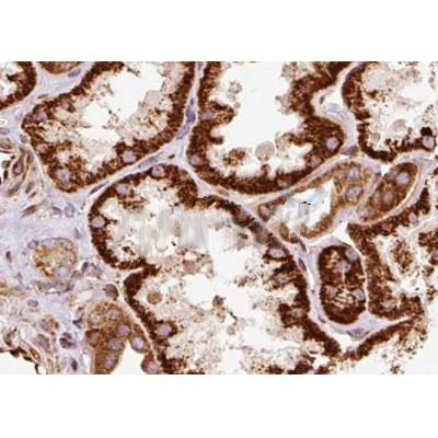
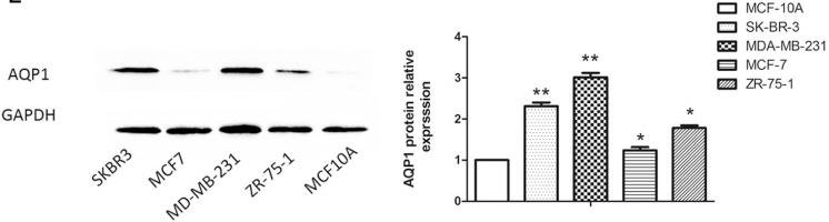
WB, IF, ICC, IHC.
Immunogen
AQP1 Antibody detects endogenous levels of total AQP1.
Reactivity
Human, Mouse, Rat.
可预测:Horse(80%), Dog(%)
Molecular weight
29kDa,35-50kDa(Glycosylation); 29kD(Calculated).
Host species
Rabbit
Ig class
Immunogen-specific rabbit IgG
Purification
Antigen affinity purification
Full name
AQP1
Synonyms
AQP 1; AQP CHIP; AQP-1; AQP1; AQP1_HUMAN; aquaporin 1 (channel-forming integral protein, 28kDa, CO blood group); aquaporin 1 (Colton blood group); Aquaporin CHIP; Aquaporin-1; Aquaporin-CHIP; Aquaporin1; Channel forming integral protein 28kDa; Channel like integral membrane protein 28 kDa; CHIP 28; CHIP28; CO; Colton blood group; Growth factor induced delayed early response protein; MGC26324; Urine water channel; Water channel protein CHIP 29; Water channel protein CHIP29; Water channel protein for red blood cells and kidney proximal tubule;
Storage
Rabbit IgG in phosphate buffered saline , pH 7.4, 150mM NaCl, 0.02% sodium azide and 50% glycerol. Store at -20 °C. Stable for 12 months from date of receipt.
Swissprot
P29972


 产品订购:
产品订购:
 渠道电话:
渠道电话: