Functions as a cell surface receptor and performs physiological functions on the surface of neurons relevant to neurite growth, neuronal adhesion and axonogenesis. Interaction between APP molecules on neighboring cells promotes synaptogenesis. Involved in cell mobility and transcription regulation through protein-protein interactions. Can promote transcription activation through binding to APBB1-KAT5 and inhibits Notch signaling through interaction with Numb. Couples to apoptosis-inducing pathways such as those mediated by G(O) and JIP. Inhibits G(o) alpha ATPase activity (By similarity). Acts as a kinesin I membrane receptor, mediating the axonal transport of beta-secretase and presenilin 1 (By similarity). By acting as a kinesin I membrane receptor, plays a role in axonal anterograde transport of cargo towards synapes in axons. Involved in copper homeostasis/oxidative stress through copper ion reduction. In vitro, copper-metallated APP induces neuronal death directly or is potentiated through Cu(2+)-mediated low-density lipoprotein oxidation. Can regulate neurite outgrowth through binding to components of the extracellular matrix such as heparin and collagen I and IV. The splice isoforms that contain the BPTI domain possess protease inhibitor activity. Induces a AGER-dependent pathway that involves activation of p38 MAPK, resulting in internalization of amyloid-beta peptide and leading to mitochondrial dysfunction in cultured cortical neurons. Provides Cu(2+) ions for GPC1 which are required for release of nitric oxide (NO) and subsequent degradation of the heparan sulfate chains on GPC1.
Amyloid-beta peptides are lipophilic metal chelators with metal-reducing activity. Bind transient metals such as copper, zinc and iron. In vitro, can reduce Cu(2+) and Fe(3+) to Cu(+) and Fe(2+), respectively. Amyloid-beta protein 42 is a more effective reductant than amyloid-beta protein 40. Amyloid-beta peptides bind to lipoproteins and apolipoproteins E and J in the CSF and to HDL particles in plasma, inhibiting metal-catalyzed oxidation of lipoproteins. APP42-beta may activate mononuclear phagocytes in the brain and elicit inflammatory responses. Promotes both tau aggregation and TPK II-mediated phosphorylation. Interaction with overexpressed HADH2 leads to oxidative stress and neurotoxicity. Also binds GPC1 in lipid rafts.
Appicans elicit adhesion of neural cells to the extracellular matrix and may regulate neurite outgrowth in the brain.
The gamma-CTF peptides as well as the caspase-cleaved peptides, including C31, are potent enhancers of neuronal apoptosis.
N-APP binds TNFRSF21 triggering caspase activation and degeneration of both neuronal cell bodies (via caspase-3) and axons (via caspase-6).
Description
Rabbit polyclonal antibody to APP
Applications
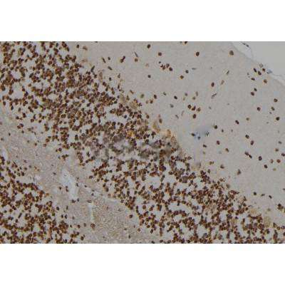
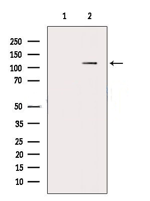
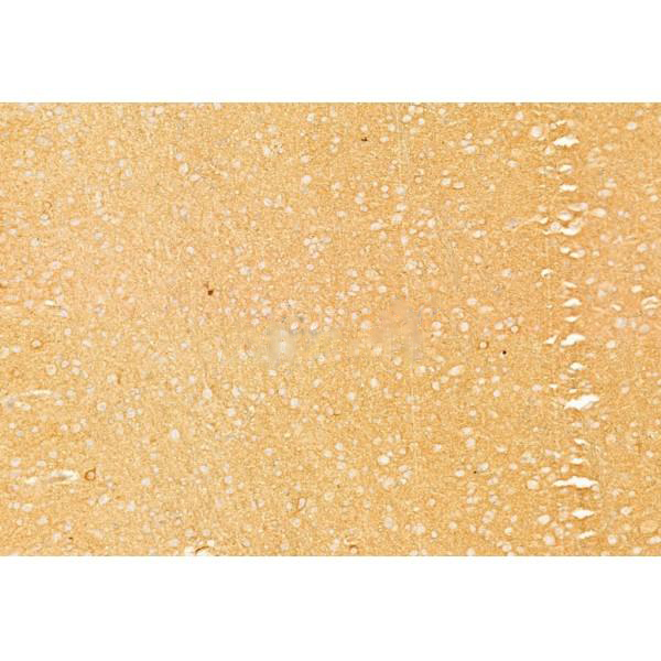
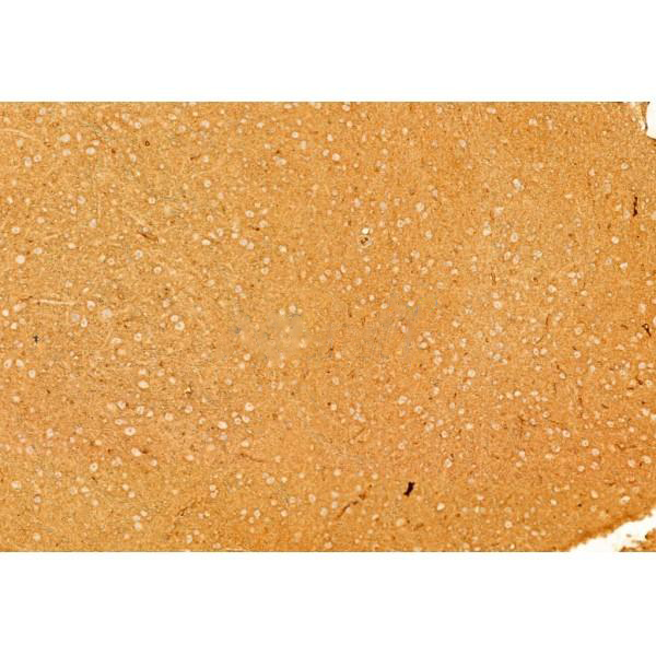
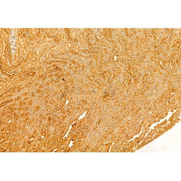
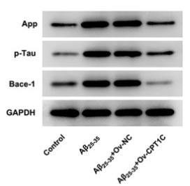
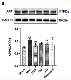
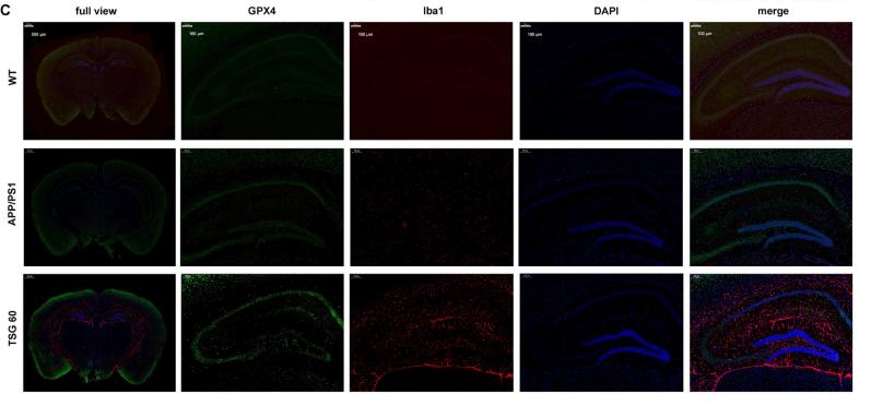
WB, IF, ICC, IHC.
Immunogen
APP Antibody detects endogenous levels of total APP.
Reactivity
Human, Mouse, Rat.
可预测:Pig(100%), Horse(%), Chicken(%), Xenopus(%)
Molecular weight
117kDa; 87kD(Calculated).
Host species
Rabbit
Ig class
Immunogen-specific rabbit IgG
Purification
Antigen affinity purification
Full name
APP
Synonyms
A4_HUMAN; AAA; ABETA; ABPP; AD1; AICD-50; AICD-57; AICD-59; AID(50); AID(57); AID(59); Alzheimer disease amyloid protein; amyloid beta A4 protein; Amyloid intracellular domain 50; Amyloid intracellular domain 57; Amyloid intracellular domain 59; amyloid of aging and alzheimer disease; APP; APPI; beta-amyloid peptide; Beta-APP40; Beta-APP42; C31; Cerebral vascular amyloid peptide; CTFgamma; CVAP; Gamma-CTF(50); Gamma-CTF(57); Gamma-CTF(59); peptidase nexin-II; PN-II; PreA4; Protease nexin-II; S-APP-alpha; S-APP-beta;
Storage
Rabbit IgG in phosphate buffered saline , pH 7.4, 150mM NaCl, 0.02% sodium azide and 50% glycerol. Store at -20 °C. Stable for 12 months from date of receipt.
Swissprot
P05067


 产品订购:
产品订购:
 渠道电话:
渠道电话: