您现在的位置:
下载说明书
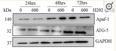
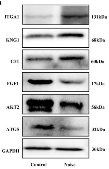
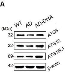
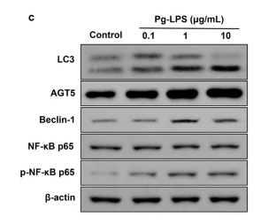
Involved in autophagic vesicle formation. Conjugation with ATG12, through a ubiquitin-like conjugating system involving ATG7 as an E1-like activating enzyme and ATG10 as an E2-like conjugating enzyme, is essential for its function. The ATG12-ATG5 conjugate acts as an E3-like enzyme which is required for lipidation of ATG8 family proteins and their association to the vesicle membranes. Involved in mitochondrial quality control after oxidative damage, and in subsequent cellular longevity. Plays a critical role in multiple aspects of lymphocyte development and is essential for both B and T lymphocyte survival and proliferation. Required for optimal processing and presentation of antigens for MHC II. Involved in the maintenance of axon morphology and membrane structures, as well as in normal adipocyte differentiation. Promotes primary ciliogenesis through removal of OFD1 from centriolar satellites and degradation of IFT20 via the autophagic pathway.
May play an important role in the apoptotic process, possibly within the modified cytoskeleton. Its expression is a relatively late event in the apoptotic process, occurring downstream of caspase activity. Plays a crucial role in IFN-gamma-induced autophagic cell death by interacting with FADD.
(Microbial infection) May act as a proviral factor. In association with ATG12, negatively regulates the innate antiviral immune response by impairing the type I IFN production pathway upon vesicular stomatitis virus (VSV) infection. Required for the translation of incoming hepatitis C virus (HCV) RNA and, thereby, for initiation of HCV replication, but not required once infection is established.
Description
Rabbit polyclonal antibody to APG5L/ATG5
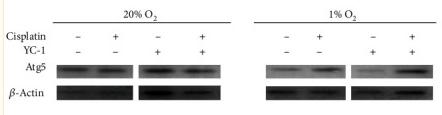
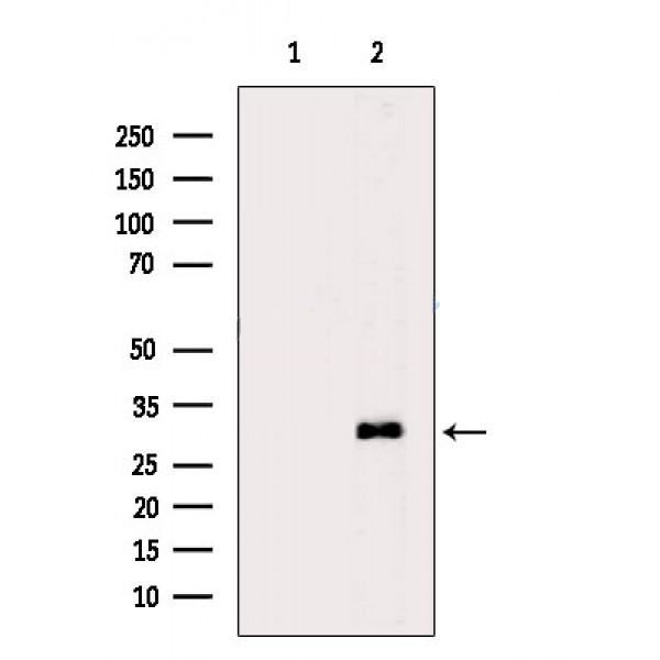
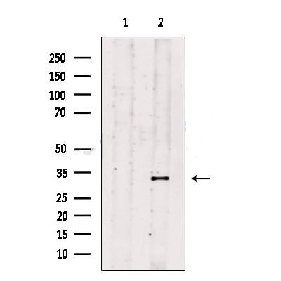
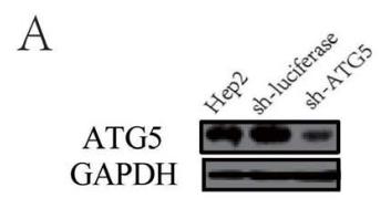
Applications
WB, IHC.
Immunogen
APG5L/ATG5 Antibody detects endogenous levels of total APG5L/ATG5.
Reactivity
Human, Mouse, Rat.
可预测:Pig(100%), Zebrafish(%), Bovine(%), Horse(%), Sheep(%), Rabbit(%), Dog(%), Chicken(%), Xenopus(%)
Molecular weight
35kD,56kD(complex); 32kD(Calculated).
Host species
Rabbit
Ig class
Immunogen-specific rabbit IgG
Purification
Antigen affinity purification
Full name
APG5L/ATG5
Synonyms
APG 5; APG 5L; APG5; APG5 autophagy 5 like; APG5 like; APG5-like; APG5L; Apoptosis specific protein; Apoptosis-specific protein; ASP; ATG 5; Atg5; ATG5 autophagy related 5 homolog; ATG5_HUMAN; Autophagy protein 5; Autophagy related 5; hAPG5; Homolog of S Cerevisiae autophagy 5; OTTHUMP00000040507;
Storage
Rabbit IgG in phosphate buffered saline , pH 7.4, 150mM NaCl, 0.02% sodium azide and 50% glycerol. Store at -20 °C. Stable for 12 months from date of receipt.
Swissprot
Q9H1Y0
Western blot analysis of extracts from Rat muscle, using APG5L/ATG5 Antibody. The lane on the left was treated with blocking peptide....
Western blot analysis of extracts from MCF7 cells, using ATG5 Antibody. The lane on the left was treated with blocking peptide....
Figure 5. |Inhibition of autophagy with specific shRNA targeting ATG5, ATG7 or BECN1 reduces RSV replication. (A-C) Hep2 cells stably expressing specific shRNA against ATG5 (Hep2-sh-ATG5), ATG7 (Hep2-sh-ATG7), BECN1 (Hep2-sh-BECN1) or luciferase (Hep2-sh-luciferase) were established as described in Materials and Methods. The silencing efficiency of ATG5 shRNA (A), ATG7 shRNA (B) and BECN1 shRNA (C) were validated by immunoblotting with anti-ATG5, anti-ATG7, anti-BECN1 and anti-GAPDH antibodies respectively....
Figure 3 Relative gene expression of apoptosis (A), autophagy (B), and necrosis (C) related genes in in-vitro primary human trabecular meshwork cells after 48 hours and 72 hours of oxidative stress with H2O2 relatively compared with control and normalized with the average CT of ACTB, GAPDH, HPRT1, RPLP20 and HGDC. (D and E) protein expression of apoptosis and autophagy markers and densitometry analysis in HTM cells after 600uM H202 treatment in HTM cells at 24, 48 and 72 hours by western. (F and G) epigenetic modifications of histones ie, H2 and H3 and densitometry analysis after 600 uM of H2O2 treatment in HTM cells at 24, 48 and 72 hours. Means ± SEM shown, *p<0.05, **p<0.01, ***p<0.001 one way ANOVA post hoc t test with Tukey correction....
Figure 5 Expression of related marker proteins and genes under normoxic and hypoxic conditions by Western blotting. (a) Beclin-1 was measured by immunoblotting under normoxia and hypoxia. (b) Caspase-3 was measured by immunoblotting when incubated with HIF-1α inhibitor YC-1. The results indicate that hypoxia might induce cell chemoresistance through the HIF-1α signaling pathway. (c) Beclin-1 was measured by immunoblotting under normoxia and hypoxia. (d) Atg5 was measured by Western blotting and showed a similar trend to caspase-3. (e) HIF-1α was measured with Western blotting. The result revealed that HIF was involved in targeting apoptosis induced by autophagy....
Figure 5 Measurement of expression levels of DEPs, oxidative stress markers and proinflammatory cytokines production. (a) Validation of representative inflammation and autophagy-related DEPs by western blots in the control group (n = 3) and noise group (n = 3). (b) Comparison of MDA level and SOD activity in cochlea between the control group (n = 3) and noise group (n = 3). (c) Comparison of proinflammatory cytokines TNF-α and IL-6 in cochlea between the control group (n = 3) and noise group (n = 3). Data are represented as mean ± SD. * p < 0.05, ** p < 0.01....
Figure 6 DHA increases the basal level of autophagy. Protein levels of ATG5, ATG12, ATG16L1, Beclin1, ATG14, p-ATG14 (Ser29), Rab7, RILP, Lamp1, CTSB, p-mTOR(Ser2448), mammalian target of rapamycin (mTOR), ULK1, p-GSK3β (Ser9), GSK3β in extracts of whole brain tissue were detected by WB. Representative Western blots are presented in panels (A,C,E,G), and quantification of the results is shown as the mean values ± SD in panels (B,D,F,H). *p < 0.05, **p < 0.01, and ***p < 0.001 between two groups. One-way analysis of variance/Newman–Keuls test was performed for all WB data....
Fig. 2 Pg-LPS treatment increased HSP90AA1 protein and gene level, and elevated the expressions of autophagy related protein in HGFs. a, b The changes of HSP90AA1 protein and gene level. c Pg-LPS (0.1, 1, 10 μg/mL) treatment increased the expression of autophagy related protein in HGFs. Data were expressed as mean ± SD, n = 3. Compared to the control group, *P ...
Figure 1. TGF-β1 induced ATG5 activation in human trabecular meshwork (HTM cells). TGF-β1 (10 ng/mL) applied to primary HTM cells for 48–72 h, increased relative mRNA expression of (A) ATG-5 and (B) LC3-II. It also increased levels of (C) ATG-5 proteins and lipidation of LC3 II, (D,E) Densitometry analysis of bands. The results represent the averages of three separate tests conducted. The error bars represent the standard deviation...

Copyright © 北京嘉美诺斯生物科技有限公司 2009. All rights reserved
备案号:湘ICP备2025088722号-1