Tumor suppressor. Promotes rapid degradation of CTNNB1 and participates in Wnt signaling as a negative regulator. APC activity is correlated with its phosphorylation state. Activates the GEF activity of SPATA13 and ARHGEF4. Plays a role in hepatocyte growth factor (HGF)-induced cell migration. Required for MMP9 up-regulation via the JNK signaling pathway in colorectal tumor cells. Acts as a mediator of ERBB2-dependent stabilization of microtubules at the cell cortex. It is required for the localization of MACF1 to the cell membrane and this localization of MACF1 is critical for its function in microtubule stabilization.
Description
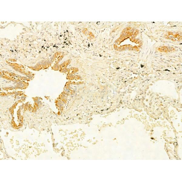
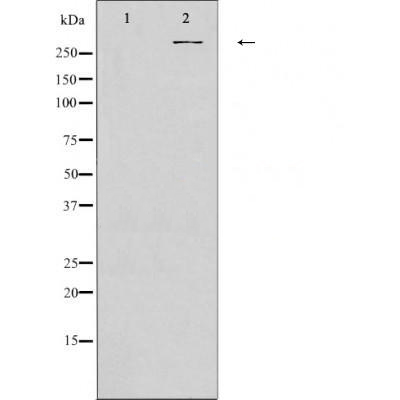
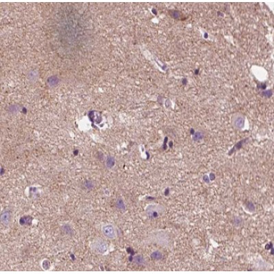
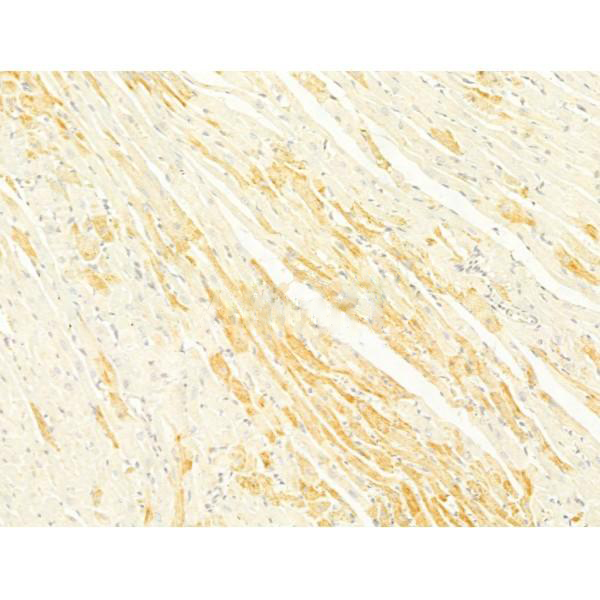
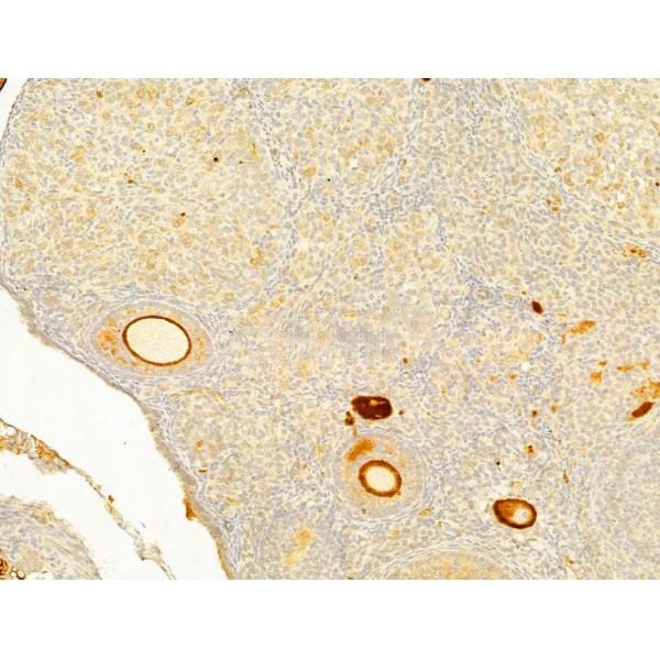
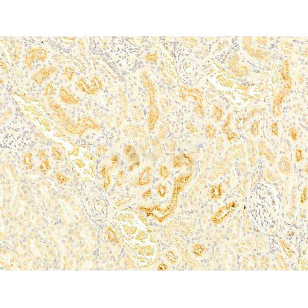
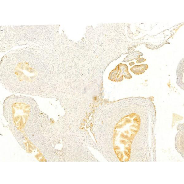
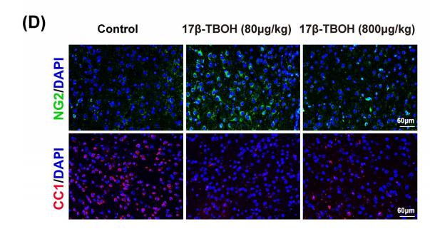
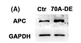
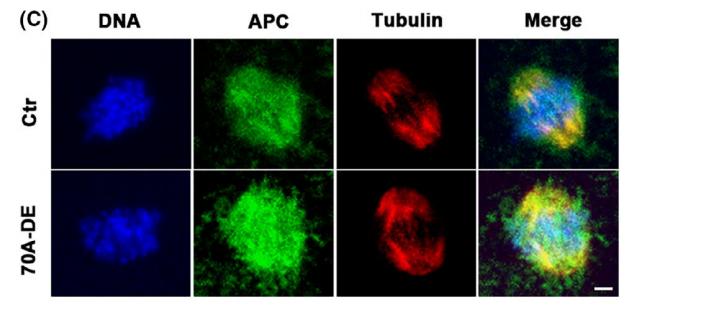
Rabbit polyclonal antibody to APC
Applications
WB, IF, ICC, IHC.
Immunogen
APC Antibody detects endogenous levels of total APC .
Reactivity
Human, Mouse, Rat.
可预测:Pig(100%), Bovine(%), Horse(%)
Molecular weight
310kDa; 312kD(Calculated).
Host species
Rabbit
Ig class
Immunogen-specific rabbit IgG
Purification
Antigen affinity purification
Full name
APC
Synonyms
Adenomatous Polyposis Coli; Adenomatous polyposis coli protein; Apc; APC_HUMAN; CC1; Deleted in polyposis 2.5; DP2; DP2.5; DP3; FAP; FPC; GS; Protein APC;
Storage
Rabbit IgG in phosphate buffered saline , pH 7.4, 150mM NaCl, 0.02% sodium azide and 50% glycerol. Store at -20 °C. Stable for 12 months from date of receipt.
Swissprot
P25054


 产品订购:
产品订购:
 渠道电话:
渠道电话: