Dioxygenase that demethylates RNA by oxidative demethylation: specifically demethylates N(6)-methyladenosine (m6A) RNA, the most prevalent internal modification of messenger RNA (mRNA) in higher eukaryotes. Can also demethylate N(6)-methyladenosine in single-stranded DNA (in vitro). Requires molecular oxygen, alpha-ketoglutarate and iron. Demethylation of m6A mRNA affects mRNA processing and export. Required for the late meiotic and haploid phases of spermatogenesis by mediating m6A demethylation in spermatocytes and round spermatids: m6A demethylation of target transcripts is required for correct splicing and the production of longer 3'-UTR mRNAs in male germ cells (By similarity).
Description
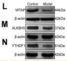
Rabbit polyclonal antibody to ALKBH5
Applications
WB, IHC.
Immunogen
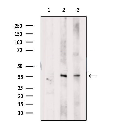
ALKBH5 Antibody detects endogenous levels of total ALKBH5.
Reactivity
Human, Mouse, Rat.
可预测:Pig(92%), Bovine(%), Horse(%), Sheep(%), Rabbit(%), Dog(%), Chicken(%)
Molecular weight
44 kDa, 37 kDa; 44kD(Calculated).
Host species
Rabbit
Ig class
Immunogen-specific rabbit IgG
Purification
Antigen affinity purification
Full name
ALKBH5
Synonyms
ABH5; AlkB, alkylation repair homolog 5 (E. coli); AlkB, alkylation repair homolog 5; Alkylated DNA repair protein alkB homolog 5; Alpha ketoglutarate dependent dioxygenase alkB homolog 5; OFOXD; OFOXD1; Oxoglutarate and iron-dependent oxygenase domain containing; Probable alpha ketoglutarate dependent dioxygenase ABH5; RNA demethylase ALKBH5;
Storage
Rabbit IgG in phosphate buffered saline , pH 7.4, 150mM NaCl, 0.02% sodium azide and 50% glycerol. Store at -20 °C. Stable for 12 months from date of receipt.
Swissprot
Q6P6C2


 产品订购:
产品订购:
 渠道电话:
渠道电话: