Serum albumin, the main protein of plasma, has a good binding capacity for water, Ca(2+), Na(+), K(+), fatty acids, hormones, bilirubin and drugs (Probable). Its main function is the regulation of the colloidal osmotic pressure of blood (Probable). Major zinc transporter in plasma, typically binds about 80% of all plasma zinc. Major calcium and magnesium transporter in plasma, binds approximately 45% of circulating calcium and magnesium in plasma (By similarity). Potentially has more than two calcium-binding sites and might additionally bind calcium in a non-specific manner (By similarity). The shared binding site between zinc and calcium at residue Asp-273 suggests a crosstalk between zinc and calcium transport in the blood (By similarity). The rank order of affinity is zinc > calcium > magnesium (By similarity). Binds to the bacterial siderophore enterobactin and inhibits enterobactin-mediated iron uptake of E.coli from ferric transferrin, and may thereby limit the utilization of iron and growth of enteric bacteria such as E.coli. Does not prevent iron uptake by the bacterial siderophore aerobactin.
Description
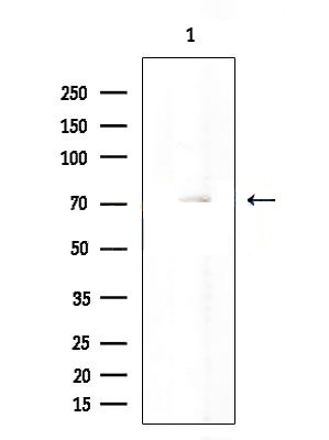
Rabbit polyclonal antibody to Albumin
Applications
WB, IHC.
Immunogen
Albumin Antibody detects endogenous levels of total Albumin.
Reactivity
Human, Mouse, Rat.
Molecular weight
69kDa; 69kD(Calculated).
Host species
Rabbit
Ig class
Immunogen-specific rabbit IgG
Purification
Antigen affinity purification
Full name
Albumin
Synonyms
alb; ALBU_HUMAN; Albumin (32 AA); Albumin (AA 34); Albumin; Analbuminemia; Bisalbuminemia; Cell growth inhibiting protein 42; DKFZp779N1935; Dysalbuminemic hyperthyroxinemia; Growth inhibiting protein 20; HSA; Hyperthyroxinemia dysalbuminemic; PRO0883; PRO0903; PRO1341; Serum albumin;
Storage
Rabbit IgG in phosphate buffered saline , pH 7.4, 150mM NaCl, 0.02% sodium azide and 50% glycerol. Store at -20 °C. Stable for 12 months from date of receipt.
Swissprot
P02768


 产品订购:
产品订购:
 渠道电话:
渠道电话: