Catalyzes the conversion of aldehydes and ketones to alcohols. Catalyzes the reduction of prostaglandin (PG) D2, PGH2 and phenanthrenequinone (PQ) and the oxidation of 9-alpha,11-beta-PGF2 to PGD2. Functions as a bi-directional 3-alpha-, 17-beta- and 20-alpha HSD. Can interconvert active androgens, estrogens and progestins with their cognate inactive metabolites. Preferentially transforms androstenedione (4-dione) to testosterone.
Description
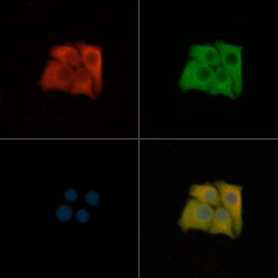
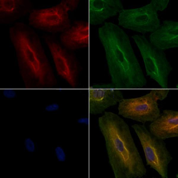
Rabbit polyclonal antibody to AKR1C3
Applications
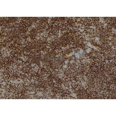
WB, IF, ICC, IHC.
Immunogen
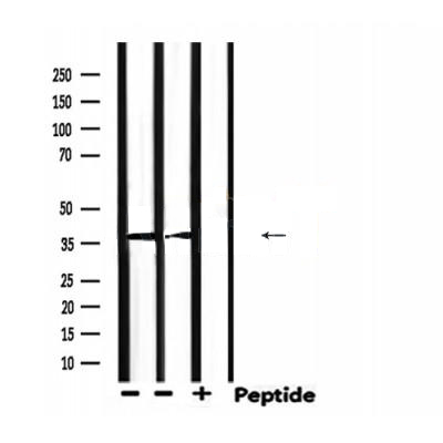
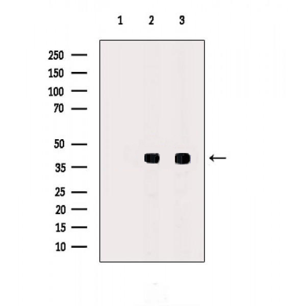
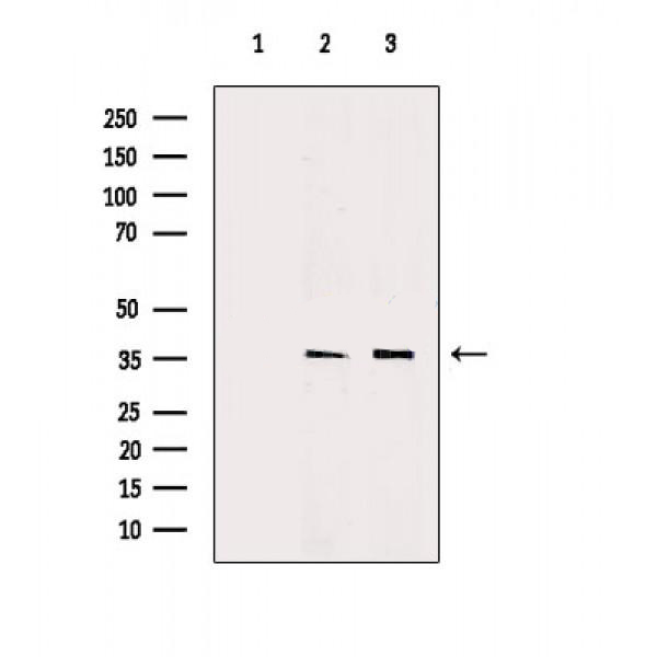
AKR1C3 Antibody detects endogenous levels of total AKR1C3.
Reactivity
Human, Mouse, Rat.
Molecular weight
37kDa; 37kD(Calculated).
Host species
Rabbit
Ig class
Immunogen-specific rabbit IgG
Purification
Antigen affinity purification
Full name
AKR1C3
Synonyms
17 beta HSD 5; 17 beta hydroxysteroid dehydrogenase type 5; 17-beta-HSD 5; 17-beta-hydroxysteroid dehydrogenase type 5; 2-dihydrobenzene-1; 2-diol dehydrogenase; 20-alpha-hydroxysteroid dehydrogenase; 3 alpha hydroxysteroid dehydrogenase type II; 3-alpha-HSD type 2; 3-alpha-HSD type II; 3-alpha-HSD type II, brain; 3-alpha-hydroxysteroid dehydrogenase type 2; AK1C3_HUMAN; AKR1 C3; Akr1c18; AKR1C3; Aldo keto reductase family 1 member C3; Aldo-keto reductase family 1 member C3; brain; Chlordecone reductase; Chlordecone reductase homolog HAKRb; DD-3; DD3; DDH1; DDX; Dihydrodiol dehydrogenase 3; Dihydrodiol dehydrogenase type I; Dihydrodiol dehydrogenase X; HA1753; HAKRB; HAKRe; hluPGFS; HSD17B5; Indanol dehydrogenase; KIAA0119; PGFS; Prostaglandin F synthase; Testosterone 17-beta-dehydrogenase 5; Trans-1; Trans-1,2-dihydrobenzene-1,2-diol dehydrogenase; Type IIb 3 alpha hydroxysteroid dehydrogenase;
Storage
Rabbit IgG in phosphate buffered saline , pH 7.4, 150mM NaCl, 0.02% sodium azide and 50% glycerol. Store at -20 °C. Stable for 12 months from date of receipt.
Swissprot
P42330


 产品订购:
产品订购:
 渠道电话:
渠道电话: