Catalyzes the reversible transfer of the terminal phosphate group between ATP and AMP. Also displays broad nucleoside diphosphate kinase activity. Plays an important role in cellular energy homeostasis and in adenine nucleotide metabolism.
Description
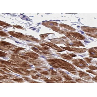
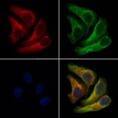
Rabbit polyclonal antibody to AK1
Applications
WB, IF, ICC, IHC.
Immunogen
AK1 Antibody detects endogenous levels of total AK1.
Reactivity
Human, Mouse, Rat.
可预测:Pig(100%), Zebrafish(%), Bovine(%), Horse(%), Sheep(%), Rabbit(%), Dog(%), Chicken(%), Xenopus(%)
Molecular weight
21kDa; 22kD(Calculated).
Host species
Rabbit
Ig class
Immunogen-specific rabbit IgG
Purification
Antigen affinity purification
Full name
AK1
Synonyms
Adenylate kinase 1; Adenylate kinase isoenzyme 1; Adenylate kinase soluble; AK 1; Ak1; ATP AMP transphosphorylase; ATP-AMP transphosphorylase 1; KAD1; KAD1_HUMAN; Myokinase;
Storage
Rabbit IgG in phosphate buffered saline , pH 7.4, 150mM NaCl, 0.02% sodium azide and 50% glycerol. Store at -20 °C. Stable for 12 months from date of receipt.
Swissprot
P00568


 产品订购:
产品订购:
 渠道电话:
渠道电话: