Ligand-activated transcriptional activator. Binds to the XRE promoter region of genes it activates. Activates the expression of multiple phase I and II xenobiotic chemical metabolizing enzyme genes (such as the CYP1A1 gene). Mediates biochemical and toxic effects of halogenated aromatic hydrocarbons. Involved in cell-cycle regulation. Likely to play an important role in the development and maturation of many tissues. Regulates the circadian clock by inhibiting the basal and circadian expression of the core circadian component PER1. Inhibits PER1 by repressing the CLOCK-ARNTL/BMAL1 heterodimer mediated transcriptional activation of PER1. The heterodimer ARNT:AHR binds to core DNA sequence 5'-TGCGTG-3' within the dioxin response element (DRE) of target gene promoters and activates their transcription.
Description
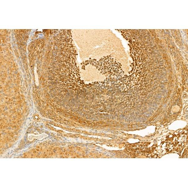
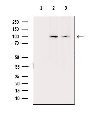
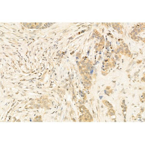
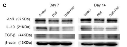
Rabbit polyclonal antibody to AhR
Applications
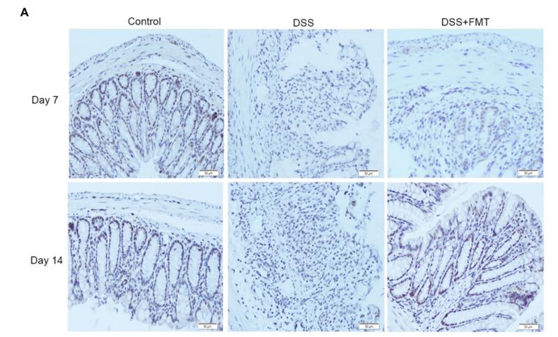
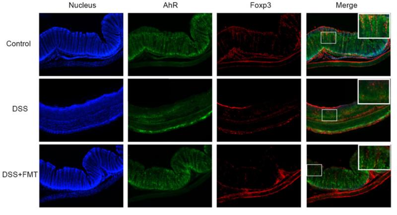
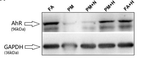
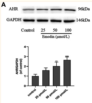
WB, IF, ICC, IHC.
Immunogen
AhR Antibody detects endogenous levels of total AhR.
Reactivity
Human, Mouse, Rat.
可预测:Pig(100%), Bovine(%), Sheep(%), Rabbit(%), Dog(%), Chicken(%), Xenopus(%)
Molecular weight
96kDa; 96kD(Calculated).
Host species
Rabbit
Ig class
Immunogen-specific rabbit IgG
Purification
Antigen affinity purification
Full name
AhR
Synonyms
Ah receptor; AhR; AHR_HUMAN; Aromatic hydrocarbon receptor; Aryl hydrocarbon receptor; Aryl hydrocarbon receptor precursor; bHLHe76; Class E basic helix loop helix protein 76; Class E basic helix-loop-helix protein 76; HGNC:348;
Storage
Rabbit IgG in phosphate buffered saline , pH 7.4, 150mM NaCl, 0.02% sodium azide and 50% glycerol. Store at -20 °C. Stable for 12 months from date of receipt.
Swissprot
P35869


 产品订购:
产品订购:
 渠道电话:
渠道电话: