heparan sulfate basal lamina glycoprotein that plays a central role in the formation and the maintenance of the neuromuscular junction (NMJ) and directs key events in postsynaptic differentiation. Component of the AGRN-LRP4 receptor complex that induces the phosphorylation and activation of MUSK. The activation of MUSK in myotubes induces the formation of NMJ by regulating different processes including the transcription of specific genes and the clustering of AChR in the postsynaptic membrane. Calcium ions are required for maximal AChR clustering. AGRN function in neurons is highly regulated by alternative splicing, glycan binding and proteolytic processing. Modulates calcium ion homeostasis in neurons, specifically by inducing an increase in cytoplasmic calcium ions. Functions differentially in the central nervous system (CNS) by inhibiting the alpha(3)-subtype of Na+/K+-ATPase and evoking depolarization at CNS synapses. This secreted isoform forms a bridge, after release from motor neurons, to basal lamina through binding laminin via the NtA domain.
transmembrane form that is the predominate form in neurons of the brain, induces dendritic filopodia and synapse formation in mature hippocampal neurons in large part due to the attached glycosaminoglycan chains and the action of Rho-family GTPases.
Isoform 1, isoform 4 and isoform 5: neuron-specific (z+) isoforms that contain C-terminal insertions of 8-19 AA are potent activators of AChR clustering. Isoform 5, agrin (z+8), containing the 8-AA insert, forms a receptor complex in myotubules containing the neuronal AGRN, the muscle-specific kinase MUSK and LRP4, a member of the LDL receptor family. The splicing factors, NOVA1 and NOVA2, regulate AGRN splicing and production of the 'z' isoforms.
Isoform 3 and isoform 6: lack any 'z' insert, are muscle-specific and may be involved in endothelial cell differentiation.
is involved in regulation of neurite outgrowth probably due to the presence of the glycosaminoglcan (GAG) side chains of heparan and chondroitin sulfate attached to the Ser/Thr- and Gly/Ser-rich regions. Also involved in modulation of growth factor signaling (By similarity).
this released fragment is important for agrin signaling and to exert a maximal dendritic filopodia-inducing effect. All 'z' splice variants (z+) of this fragment also show an increase in the number of filopodia.
Description
Rabbit polyclonal antibody to AGRN
Applications
WB, IF, ICC.
Immunogen
AGRN Antibody detects endogenous levels of total AGRN.
Reactivity
Human, Mouse, Rat.
可预测:Pig(100%), Zebrafish(%), Bovine(%), Dog(%), Chicken(%)
Molecular weight
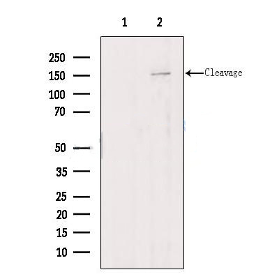
217kDa, 150 kDa; 217kD(Calculated).
Host species
Rabbit
Ig class
Immunogen-specific rabbit IgG
Purification
Antigen affinity purification
Full name
AGRN
Synonyms
AGRIN; Agrin proteoglycan; AGRN; FLJ45064; OTTHUMP00000044043;
Storage
Rabbit IgG in phosphate buffered saline , pH 7.4, 150mM NaCl, 0.02% sodium azide and 50% glycerol. Store at -20 °C. Stable for 12 months from date of receipt.
Swissprot
O00468


 产品订购:
产品订购:
 渠道电话:
渠道电话: