This proteoglycan is a major component of extracellular matrix of cartilagenous tissues. A major function of this protein is to resist compression in cartilage. It binds avidly to hyaluronic acid via an N-terminal globular region.
Description
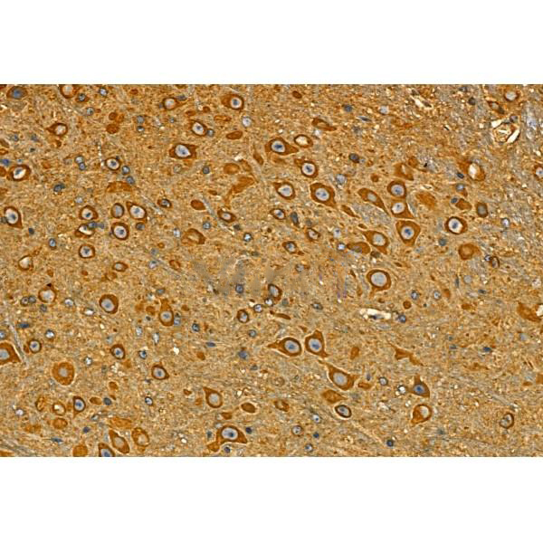
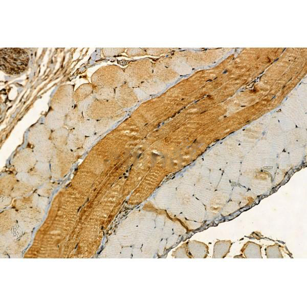
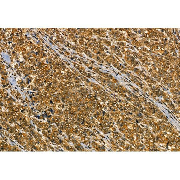
Rabbit polyclonal antibody to Aggrecan
Applications
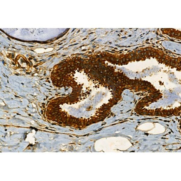
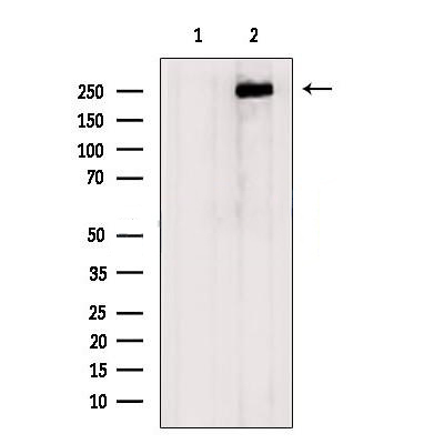
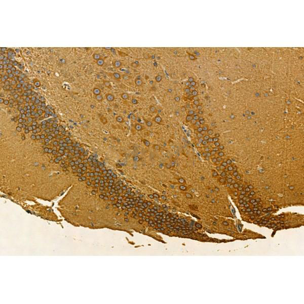
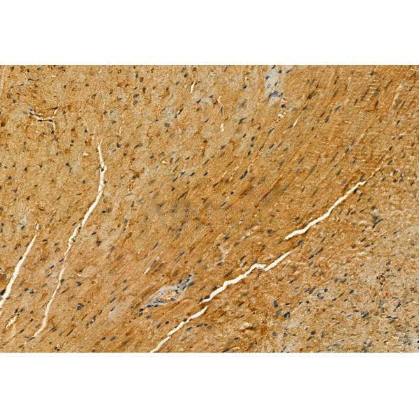
WB, IF, ICC, IHC.
Immunogen
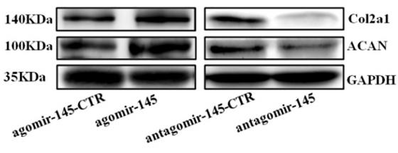
Aggrecan Antibody detects endogenous levels of total Aggrecan.
Reactivity
Human, Mouse, Rat.
可预测:Pig(100%), Bovine(%), Sheep(%), Rabbit(%), Dog(%)
Molecular weight
70,150,250 kDa; 261kD(Calculated).
Host species
Rabbit
Ig class
Immunogen-specific rabbit IgG
Purification
Antigen affinity purification
Full name
Aggrecan
Synonyms
ACAN; AGC 1; AGC1; AGCAN; Aggrecan 1 (chondroitin sulfate proteoglycan 1, large aggregating proteoglycan, antigen identified by monoclonal antibody A0122); Aggrecan 1; Aggrecan core protein; Aggrecan proteoglycan; Aggrecan structural proteoglycan of cartilage; Aggrecan1; ATEGQV; Cartilage specific proteoglycan core protein; Chondroitin sulfate proteoglycan 1; Chondroitin sulfate proteoglycan 1 large aggregating proteoglycan antigen identified by monoclonal antibody A0122; Chondroitin sulfate proteoglycan core protein 1; CSPG 1; CSPG1; CSPGCP; JSCATE; Large aggregating proteoglycan; mcspg; mgsk16; MSK 16; MSK16; SEDK;
Storage
Rabbit IgG in phosphate buffered saline , pH 7.4, 150mM NaCl, 0.02% sodium azide and 50% glycerol. Store at -20 °C. Stable for 12 months from date of receipt.
Swissprot
P16112


 产品订购:
产品订购:
 渠道电话:
渠道电话: