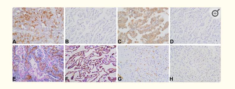
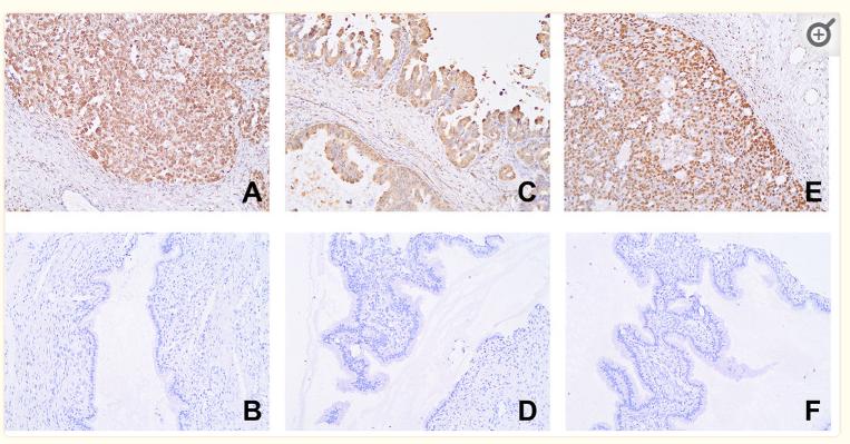
Promotes angiogenesis and the proliferation of endothelial cells. Able to bind to endothelial cells and promote cell proliferation, suggesting that it may act in an autocrine fashion.
Description
Rabbit polyclonal antibody to AGGF1
Applications
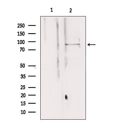
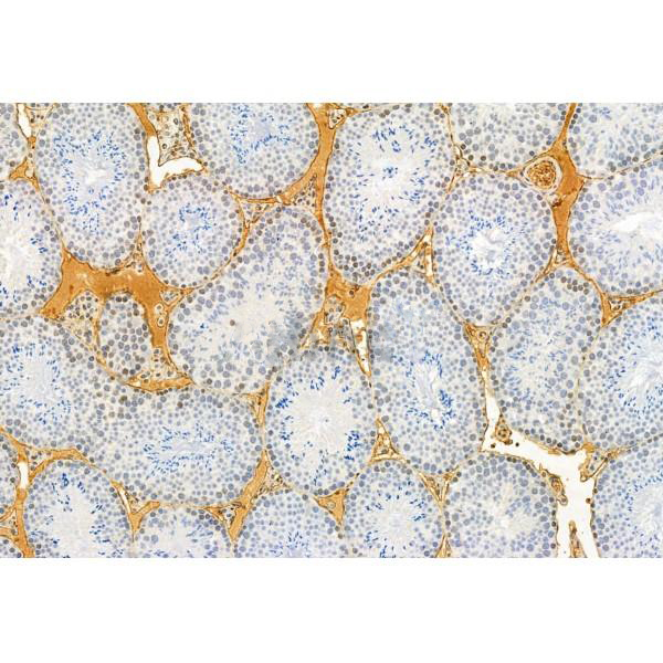
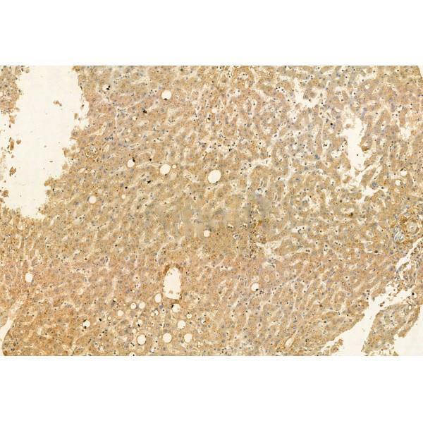
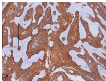
WB, IHC.
Immunogen
AGGF1 Antibody detects endogenous levels of total AGGF1.
Reactivity
Human, Mouse, Rat.
可预测:Pig(100%), Zebrafish(%), Bovine(%), Horse(%), Sheep(%), Dog(%), Chicken(%), Xenopus(%)
Molecular weight
84-100 kDa; 81kD(Calculated).
Host species
Rabbit
Ig class
Immunogen-specific rabbit IgG
Purification
Antigen affinity purification
Full name
AGGF1
Synonyms
AGGF 1; Aggf1; AGGF1_HUMAN; Angiogenic factor VG5Q; Angiogenic factor with G patch and FHA domains 1; G patch domain containing protein 7; G patch domain-containing protein 7; GPATC 7; GPATC7; GPATCH 7; GPATCH7; HSU84971; HUS84971; hVG5Q; Vasculogenesis gene on 5q; Vasculogenesis gene on 5q protein; VG5Q;
Storage
Rabbit IgG in phosphate buffered saline , pH 7.4, 150mM NaCl, 0.02% sodium azide and 50% glycerol. Store at -20 °C. Stable for 12 months from date of receipt.
Swissprot
Q8N302


 产品订购:
产品订购:
 渠道电话:
渠道电话: