On ligand binding, forms a receptor complex consisting of two type II and two type I transmembrane serine/threonine kinases. Type II receptors phosphorylate and activate type I receptors which autophosphorylate, then bind and activate SMAD transcriptional regulators. Receptor for activin A, activin B and inhibin A. Mediates induction of adipogenesis by GDF6 (By similarity).
Description
Rabbit polyclonal antibody to ACVR2A
Applications
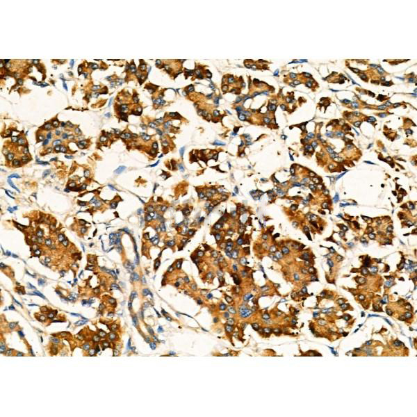
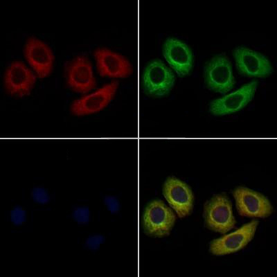
WB, IF, ICC, IHC
Immunogen
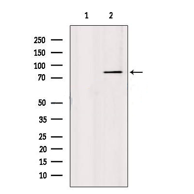
ACVR2A Antibody detects endogenous levels of total ACVR2A.
Reactivity
Human, Mouse, Rat.
可预测:Pig(100%), Bovine(100%), Horse(100%), Sheep(100%), Rabbit(100%), Dog(100%), Chicken(100%), Xenopus(100%)
Molecular weight
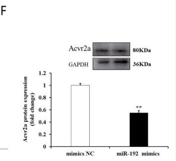
57kDa, 80 kDa; 58kD(Calculated).
Host species
Rabbit
Ig class
Immunogen-specific rabbit IgG
Purification
Antigen affinity purification
Full name
ACVR2A
Synonyms
Activin A receptor type IIA;Activin receptor type 2A;Activin receptor type IIA;Activin receptor type-2A;ACTR 2;ACTR IIA;ACTR-IIA;ACTR2;ACTRII;ACTRIIA;Acvr 2;Acvr 2A;Acvr2;ACVR2A;AVR2A_HUMAN;OTTHUMP00000197918;
Storage
Rabbit IgG in phosphate buffered saline , pH 7.4, 150mM NaCl, 0.02% sodium azide and 50% glycerol. Store at -20 °C. Stable for 12 months from date of receipt.
Swissprot
P27037


 产品订购:
产品订购:
 渠道电话:
渠道电话: