Catalyzes the conversion of long-chain fatty acids to their active form acyl-CoA for both synthesis of cellular lipids, and degradation via beta-oxidation. Preferentially activates arachidonate and eicosapentaenoate as substrates. Preferentially activates 8,9-EET > 14,15-EET > 5,6-EET > 11,12-EET. Modulates glucose-stimulated insulin secretion by regulating the levels of unesterified EETs (By similarity). Modulates prostaglandin E2 secretion.
Description
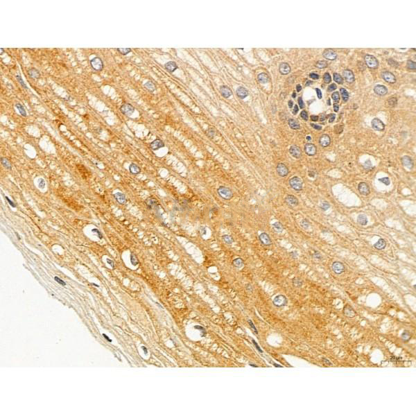
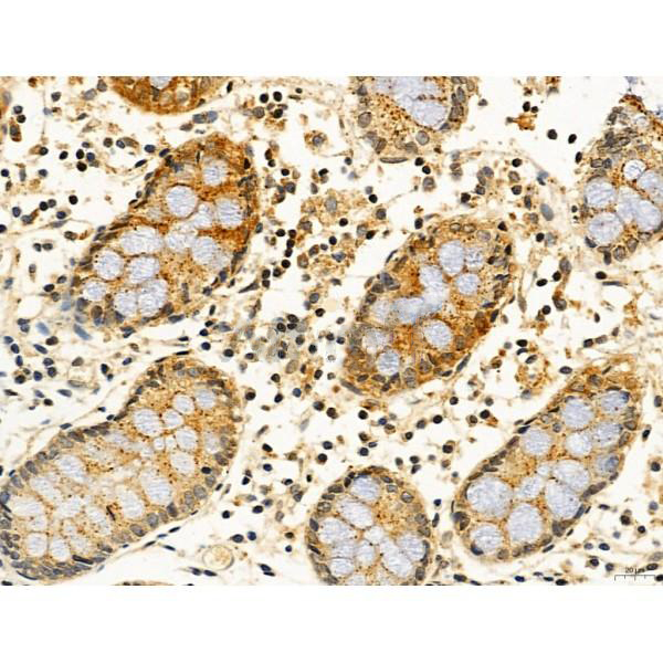
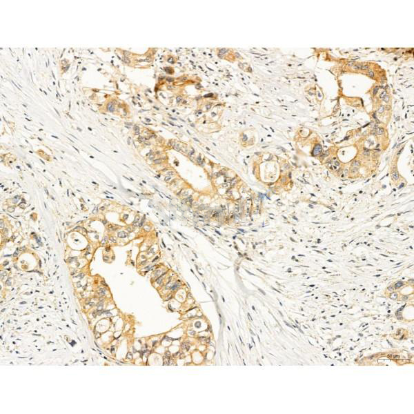
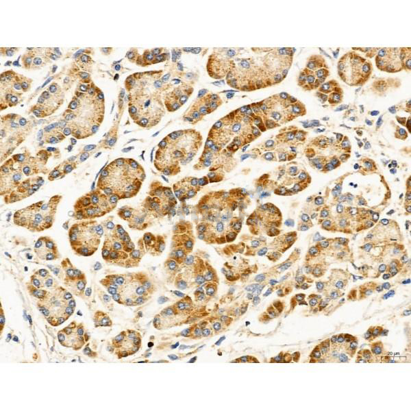
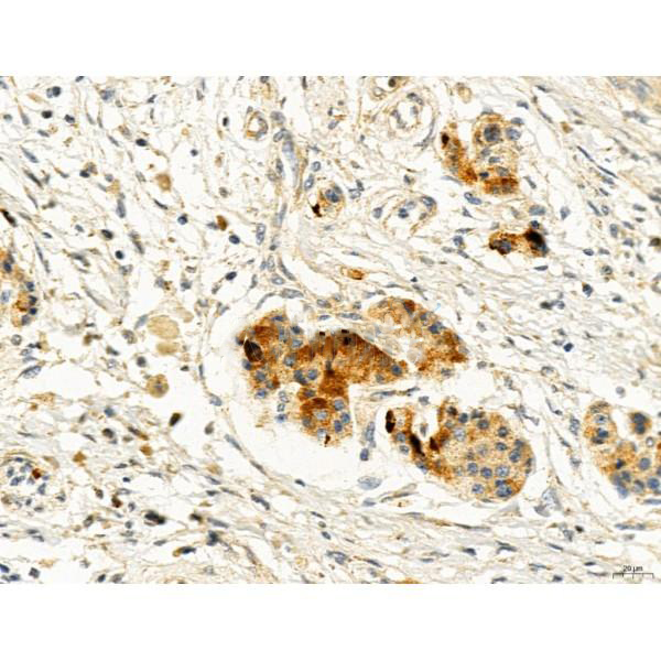
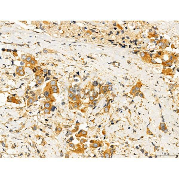
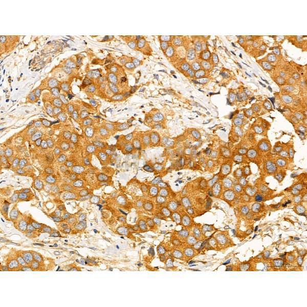
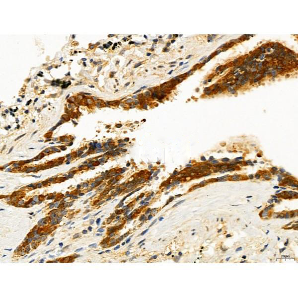
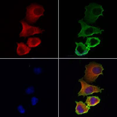
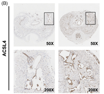
Rabbit polyclonal antibody to ACSL4/FACL4
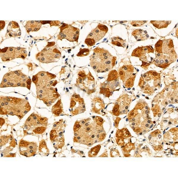
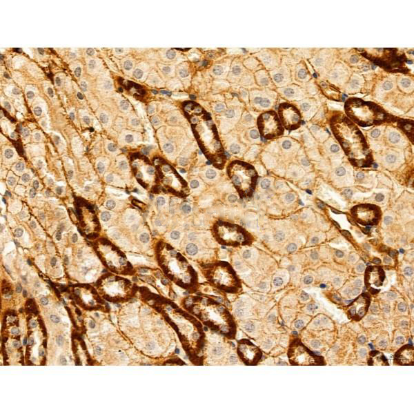
Applications
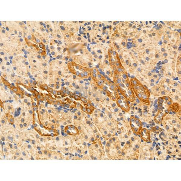
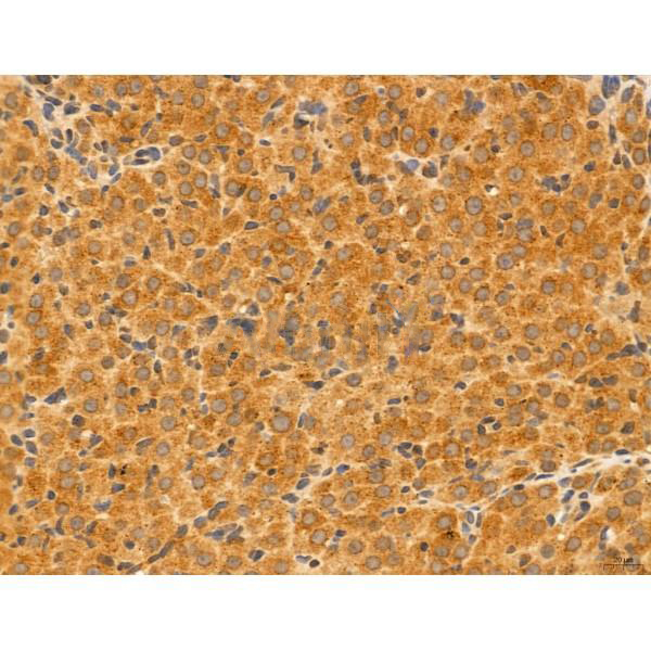
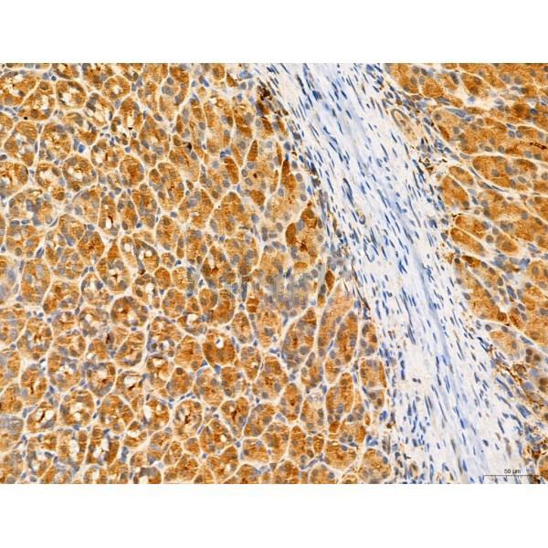
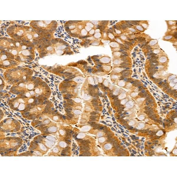
WB, IF, ICC, IHC
Immunogen
ACSL4/FACL4 Antibody detects endogenous levels of total ACSL4/FACL4.
Reactivity
Human, Mouse, Rat.
可预测:Pig(100%), Bovine(100%), Horse(100%), Sheep(100%), Rabbit(100%), Dog(100%), Chicken(100%), Xenopus(83%)
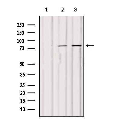
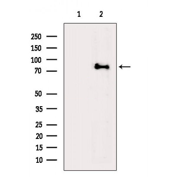
Molecular weight
79 kDa,74 kDa; 79kD(Calculated).
Host species
Rabbit
Ig class
Immunogen-specific rabbit IgG
Purification
Antigen affinity purification
Full name
ACSL4/FACL4
Synonyms
ACS 4; ACS4; ACSL 4; Acsl4; ACSL4_HUMAN; acyl CoA synthetase 4; Acyl CoA synthetase long chain family member 4; FACL 4; FACL4; Fatty acid Coenzyme A ligase; fatty acid Coenzyme A ligase long-chain 4; LACS 4; LACS4; Lignoceroyl CoA synthase; Long chain 4; long chain acyl CoA synthetase 4; long chain fatty acid CoA ligase 4; long chain fatty acid Coenzyme A ligase 4; Long-chain acyl-CoA synthetase 4; Long-chain-fatty-acid--CoA ligase 4; MRX63; MRX68;
Storage
Rabbit IgG in phosphate buffered saline , pH 7.4, 150mM NaCl, 0.02% sodium azide and 50% glycerol. Store at -20 °C. Stable for 12 months from date of receipt.
Swissprot
O60488


 产品订购:
产品订购:
 渠道电话:
渠道电话: