Broad substrate specificity ATP-dependent transporter of the ATP-binding cassette (ABC) family that actively extrudes a wide variety of physiological compounds, dietary toxins and xenobiotics from cells. Involved in porphyrin homeostasis, mediating the export of protoporphyrin IX (PPIX) from both mitochondria to cytosol and cytosol to extracellular space, it also functions in the cellular export of heme. Also mediates the efflux of sphingosine-1-P from cells. Acts as a urate exporter functioning in both renal and extrarenal urate excretion. In kidney, it also functions as a physiological exporter of the uremic toxin indoxyl sulfate (By similarity). Also involved in the excretion of steroids like estrone 3-sulfate/E1S, 3beta-sulfooxy-androst-5-en-17-one/DHEAS, and other sulfate conjugates. Mediates the secretion of the riboflavin and biotin vitamins into milk (By similarity). Extrudes pheophorbide a, a phototoxic porphyrin catabolite of chlorophyll, reducing its bioavailability (By similarity). Plays an important role in the exclusion of xenobiotics from the brain (Probable). It confers to cells a resistance to multiple drugs and other xenobiotics including mitoxantrone, pheophorbide, camptothecin, methotrexate, azidothymidine, and the anthracyclines daunorubicin and doxorubicin, through the control of their efflux. In placenta, it limits the penetration of drugs from the maternal plasma into the fetus (By similarity). May play a role in early stem cell self-renewal by blocking differentiation (By similarity).
Description
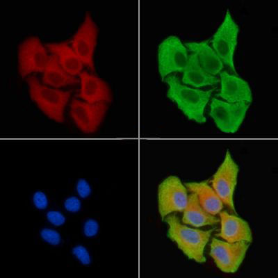
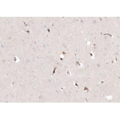
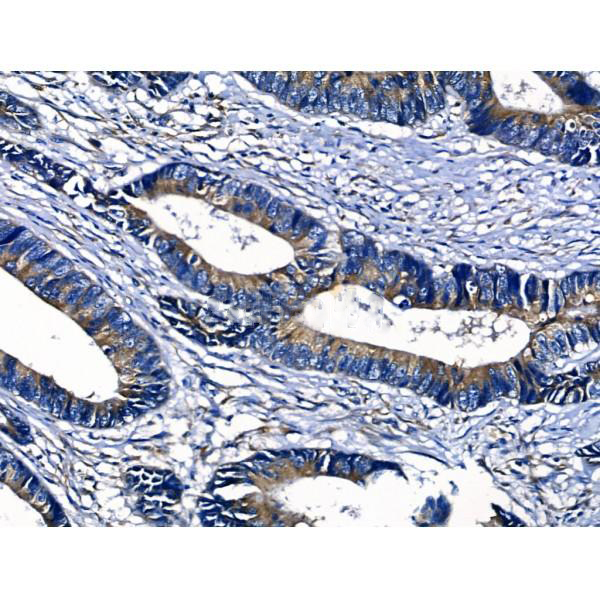
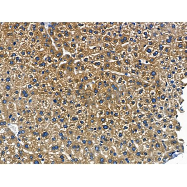
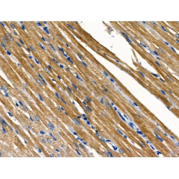
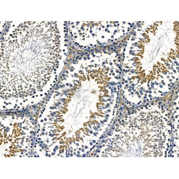
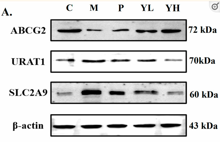
Rabbit polyclonal antibody to ABCG2
Applications
WB, IF, ICC, IHC
Immunogen
ABCG2 Antibody detects endogenous levels of total ABCG2.
Reactivity
Human, Mouse, Rat.
可预测:Rabbit(89%)
Molecular weight
72 kDa; 72kD(Calculated).
Host species
Rabbit
Ig class
Immunogen-specific rabbit IgG
Purification
Antigen affinity purification
Full name
ABCG2
Synonyms
ABC transporter; ABC15; ABCG 2; ABCG2; ABCG2_HUMAN; ABCP; ATP binding cassette sub family G (WHITE) member 2; ATP binding cassette transporter G2; ATP-binding cassette sub-family G member 2; BCRP; BCRP1; BMDP; Breast cancer resistance protein; CD338; CDw338; CDw338 antigen; EST157481; GOUT1; MGC102821; Mitoxantrone resistance associated protein; Mitoxantrone resistance-associated protein; MRX; Multi drug resistance efflux transport ATP binding cassette sub family G (WHITE) member 2; MXR; MXR1; Placenta specific ATP binding cassette transporter; Placenta specific MDR protein; Placenta-specific ATP-binding cassette transporter; UAQTL1;
Storage
Rabbit IgG in phosphate buffered saline , pH 7.4, 150mM NaCl, 0.02% sodium azide and 50% glycerol. Store at -20 °C. Stable for 12 months from date of receipt.
Swissprot
Q9UNQ0


 产品订购:
产品订购:
 渠道电话:
渠道电话: