Double-strand break (DSB) repair protein involved in response to DNA damage, telomere dynamics and class-switch recombination (CSR) during antibody genesis. Plays a key role in the repair of double-strand DNA breaks (DSBs) in response to DNA damage by promoting non-homologous end joining (NHEJ)-mediated repair of DSBs and specifically counteracting the function of the homologous recombination (HR) repair protein BRCA1. In response to DSBs, phosphorylation by ATM promotes interaction with RIF1 and dissociation from NUDT16L1/TIRR, leading to recruitment to DSBs sites. Recruited to DSBs sites by recognizing and binding histone H2A monoubiquitinated at 'Lys-15' (H2AK15Ub) and histone H4 dimethylated at 'Lys-20' (H4K20me2), two histone marks that are present at DSBs sites. Required for immunoglobulin class-switch recombination (CSR) during antibody genesis, a process that involves the generation of DNA DSBs. Participates to the repair and the orientation of the broken DNA ends during CSR (By similarity). In contrast, it is not required for classic NHEJ and V(D)J recombination (By similarity). Promotes NHEJ of dysfunctional telomeres via interaction with PAXIP1.
Description
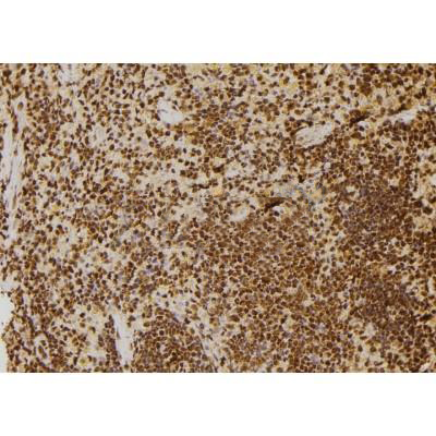
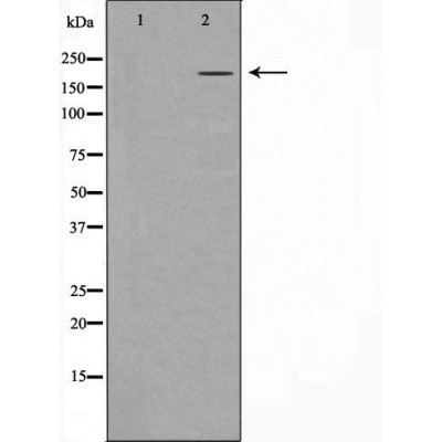
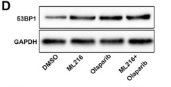
Rabbit polyclonal antibody to 53BP1
Applications
WB, IF, ICC, IHC
Immunogen
53BP1 Antibody detects endogenous levels of total 53BP1.
Reactivity
Human, Mouse, Rat.
可预测:Pig(100%), Horse(100%), Sheep(100%), Rabbit(100%), Dog(100%)
Molecular weight
213kDa; 214kD(Calculated).
Host species
Rabbit
Ig class
Immunogen-specific rabbit IgG
Purification
Antigen affinity purification
Full name
53BP1
Synonyms
53 BP1; 53BP1; FLJ41424; MGC138366; p202; p53 binding protein 1; p53 BP1; p53-binding protein 1; p53BP1; TP53 BP1; TP53B_HUMAN; Tp53bp1; TRP53 BP1; Tumor protein 53 binding protein 1; Tumor protein p53 binding protein 1; Tumor suppressor p53 binding protein 1; Tumor suppressor p53-binding protein 1;
Storage
Rabbit IgG in phosphate buffered saline , pH 7.4, 150mM NaCl, 0.02% sodium azide and 50% glycerol. Store at -20 °C. Stable for 12 months from date of receipt.
Swissprot
Q12888


 产品订购:
产品订购:
 渠道电话:
渠道电话: